Welcome to the Amira-Avizo Software Use Case Gallery
Below you will find a collection of use cases of our 3D data visualization and analysis software. These use cases include scientific publications, articles, papers, posters, presentations or even videos that show how Amira-Avizo Software is used to address various scientific and industrial research topics.
Use the Domain selector to filter by main application area, and use the Search box to enter keywords related to specific topics you are interested in.
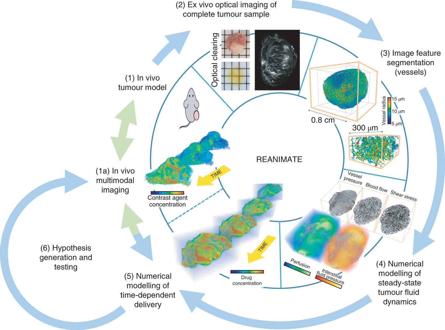
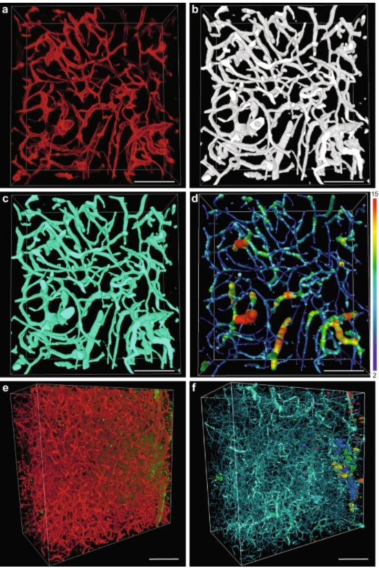
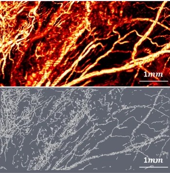
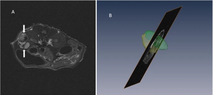
Understanding the uptake of a drug by diseased tissue, and the drug’s subsequent spatiotemporal distribution, are central factors in the development of effective targeted therapies. However, the interaction between the pathophysiology of diseased tissue and individual therapeutic agents can be complex, and can vary across tissue types and across subjects. Here, we show that the combination of mathematical modelling, high-resolution optical imaging of intact and optically cleared tumour tiss... Read more
Angela d’Esposito, Paul W. Sweeney, Morium Ali, Magdy Saleh, Rajiv Ramasawmy, Thomas A. Roberts, Giulia Agliardi, Adrien Desjardins, Mark F. Lythgoe, R. Barbara Pedley, Rebecca Shipley and Simon Walker-Samuel
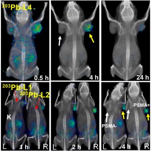
Targeted radiopharmaceutical therapy (TRT) using α-particle radiation is a promising approach for treating both large and micrometastatic lesions. Researchers developed prostate-specific membrane antigen (PSMA)–targeted low-molecular-weight agents for 212Pb-based TRT of patients with prostate cancer (PC) by evaluating the matching γ-emitting surrogate.
Targeted radiopharmaceutical therapy using α-particles (α-TRTs), which cause deposition of ionizing radiation of high-linear-ener... Read more
Sangeeta Ray Banerjee, Il Minn, Vivek Kumar, Anders Josefsson, Ala Lisok, Mary Brummet, Jian Chen, Ana P. Kiess, Kwamena Baidoo, Cory Brayton, Ronnie C. Mease, Martin Brechbiel, George Sgouros, Robert F. Hobbs, and Martin G. Pomper
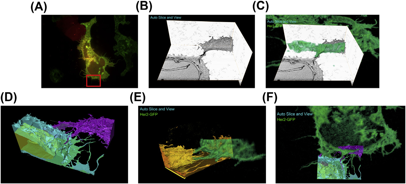
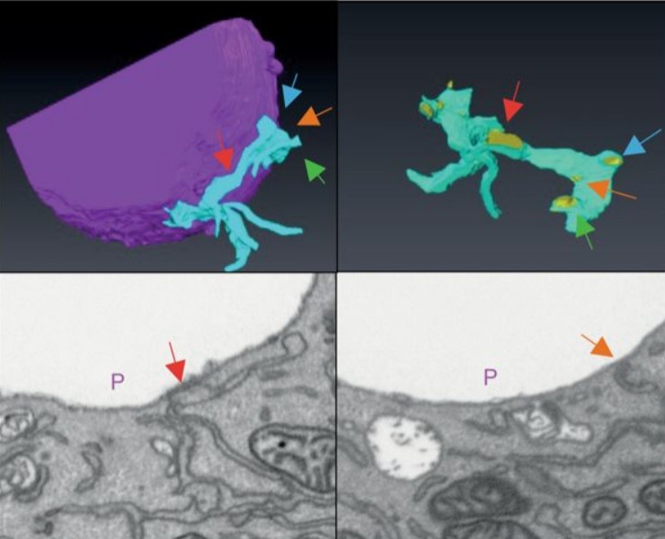
A fully integrated, three-dimensional fluorescence to electron microscopy correlative workflow
While fluorescence microscopy provides tools for highly specific labeling and sensitive detection, its resolution limit and lack of general contrast has hindered studies of cellular structure and protein localization. Recent advances in correlative light and electron microscopy (CLEM), including the fully integrated CLEM workflow instrument, the Thermo Scientific CorrSight with MAPS, have allowed for a more reliable, reproducible, and quicker approach to correlate three-dimensional time-lapse... Read more
Claudia S. Lopez, Cedric Bouchet-Marquis, Christopher P. Arthur, Jessica L. Riesterer, Gregor Heiss, Guillaume Thibault, Lee Pullan, Sunjong Kwon, Joe W. Gray
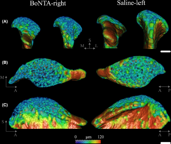
Masseter muscle function influences mandibular bone homeostasis. As previously reported, bone resorption markers increased in the mouse mandibular condyle two days after masseter paralysis induced with botulinum toxin type A (BoNTA), followed by local bone loss.
This study aimed to evaluate the bone quality of both the mandibular condyle and alveolar process in the mandible of adult mice during the early stage of a BoNTA‐induced masseter muscle atrophy, using a combined 3D histomorpho... Read more
Julián Balanta‐Melo, María Angélica Torres‐Quintana, Maximilian Bemmann, Carolina Vega, Constanza González, Kornelius Kupczik, Viviana Toro‐Ibacache, Sonja Buvinic

Protocols for Generating Surfaces and Measuring 3D Organelle Morphology Using Amira
High-resolution 3D images of organelles are of paramount importance in cellular biology. Although light microscopy and transmission electron microscopy (TEM) have provided the standard for imaging cellular structures, they cannot provide 3D images.
However, recent technological advances such as serial block-face scanning electron microscopy (SBF-SEM) and focused ion beam scanning electron microscopy (FIB-SEM) provide the tools to create 3D images for the ultrastructural analysis of org... Read more
Edgar Garza-Lopez, Zer Vue, Prasanna Katti, Kit Neikirk, Michelle Biete, Jacob Lam, Heather K. Beasley, Andrea G. Marshall, Taylor A. Rodman, Trace A. Christensen, Jeffrey L. Salisbury, Larry Vang, Margaret Mungai, Salma Ash Shareef, Sandra A. Murray, Jianqiang Shao, Jennifer Streeter, Brian Glancy, Renata O. Pereira1, E. Dale Abel, and Antentor Hinton, Jr.
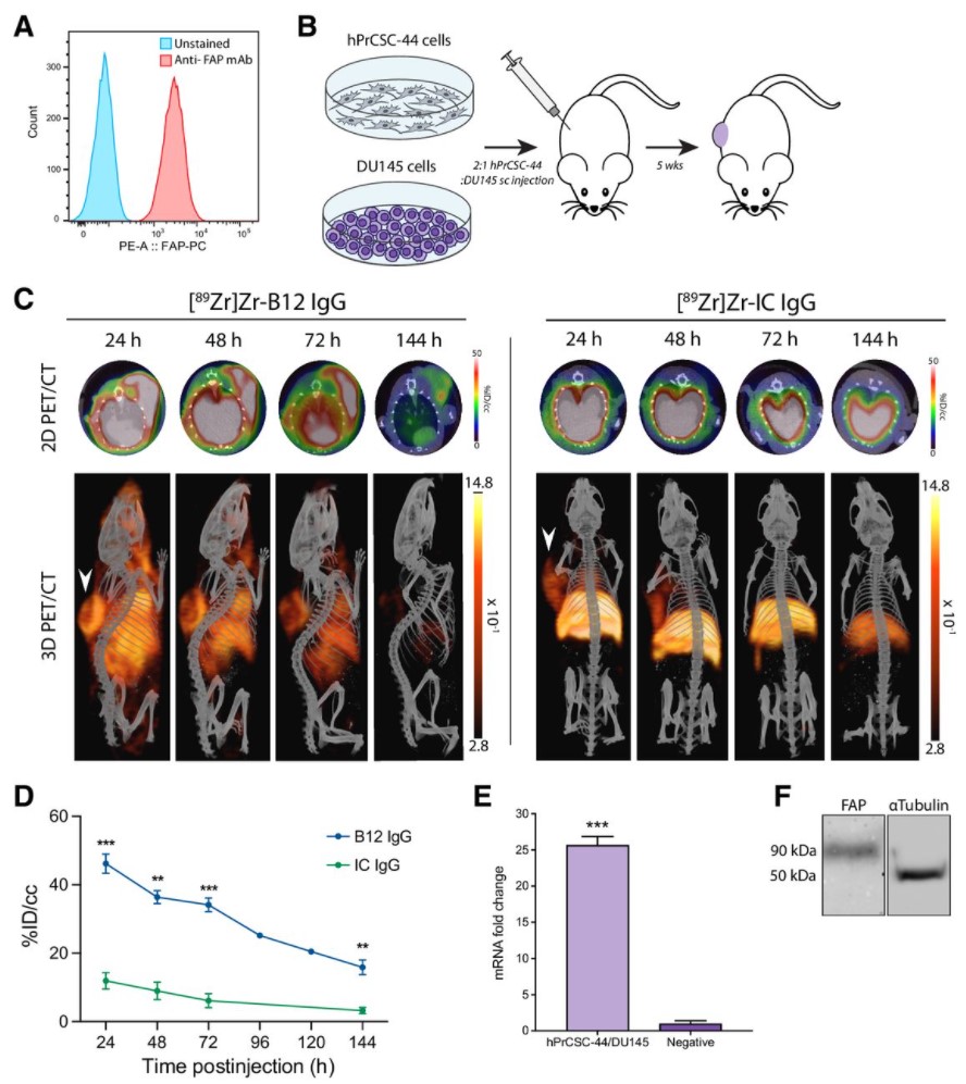
Malignant cells are surrounded by a complex and supportive tumor microenvironment that consists of immune cells, extracellular matrix, vasculature, and fibroblasts. Cancer-associated fibroblasts (CAF) are the major cell type in the reactive stroma and are known to promote tumorigenesis and metastasis. Fibroblast activation protein alpha (FAP) is a transmembrane serine protease expressed by CAFs in the microenvironment of epithelial tumors.
Meta-analysis of FAP expression and clinical ... Read more
Hallie M. Hintz, Joseph P. Gallant, Donald J. Vander Griend, Ilsa M. Coleman, Peter S. Nelson and Aaron M. LeBeau
Precise methods for quantifying drug accumulation in brain tissue are currently very limited, challenging the development of new therapeutics for brain disorders. Transcardial perfusion is instrumental for removing the intravascular fraction of an injected compound, thereby allowing for ex vivo assessment of extravasation into the brain. However, pathological remodeling of tissue microenvironment can affect the efficiency of transcardial perfusion, which has been largely overlooked.
We... Read more
Serhii Kostrikov, Kasper B. Johnsen, Thomas H. Braunstein, Johann M. Gudbergsson, Frederikke P. Fliedner, Elisabeth A. A. Obara, Petra Hamerlik, Anders E. Hansen, Andreas Kjaer, Casper Hempel & Thomas L. Andresen
Optical Resolution Photoacoustic Microscopy of Ovary and Fallopian Tube
Ovarian cancer is the leading cause of death among gynecological cancers, but is poorly amenable to preoperative diagnosis. In this study, we investigate the feasibility of “optical biopsy,” using high-optical-resolution photoacoustic microscopy (OR-PAM) to quantify the microvasculature of ovarian and fallopian tube tissue. The technique is demonstrated using excised human ovary and fallopian tube specimens imaged immediately after surgery.
This report describes the first applicatio... Read more
Bin Rao, Xiandong Leng, Yifeng Zeng, Yixiao Lin, Ruimin Chen, Qifa Zhou, Andrea R. Hagemann, Lindsay M. Kuroki, Carolyn K. McCourt, David G. Mutch, Matthew A. Powell, Ian S. Hagemann & Quing Zhu
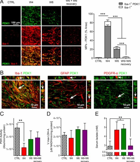
Proinflammatory mononuclear phagocytes (MPs) play a crucial role in the progression of multiple sclerosis (MS) and other neurodegenerative diseases. Despite advances in neuroimaging, there are currently limited available methods enabling noninvasive detection of MPs in vivo. Interestingly, upon activation and subsequent differentiation toward a proinflammatory phenotype MPs undergo metabolic reprogramming that results in increased glycolysis and production of lactate. Hyperpolarized (HP)
Caroline Guglielmetti, Chloé Najac, Alessandro Didonna, Annemie Van der Linden, Sabrina M. Ronen, and Myriam M. Chaumeil
Recent treatment developments for metastatic renal cell carcinoma offer combinations of immunotherapies or immunotherapy associated with tyrosine kinase inhibitors (TKI). There is currently no argument to choose one solution or another. Easy-to-use markers to assess longitudinal responses to TKI are necessary to determine when to switch to immunotherapies. These new markers will enable an earlier adaptation of therapeutic strategy in order to prevent tumor development, unnecessary toxicity an... Read more
Alexandre Ingels, Ingrid Leguerney, Paul-Henry Cournède, Jacques Irani, Sophie Ferlicot, Catherine Sébrié, Baya Benatsou, Laurène Jourdain, Stephanie Pitre-Champagnat, Jean-Jacques Patard & Nathalie Lassau
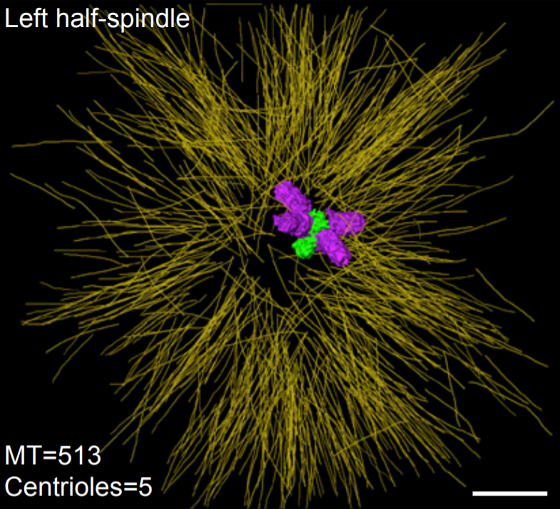
Asymmetric Centriole Numbers at Spindle Poles Cause Chromosome Missegregation in Cancer
Chromosomal instability is a hallmark of cancer and correlates with the presence of extra centrosomes, which originate from centriole overduplication.
Overduplicated centrioles lead to the formation of centriole rosettes, which mature into supernumerary centrosomes in the subsequent cell cycle. While extra centrosomes promote chromosome missegregation by clustering into pseudo-bipolar spindles, the contribution of centriole rosettes to chromosome missegregation is unknown. We us... Read more
Marco R.Cosenza, Anna Cazzola, Annik Rossberg, Nicole L. Schieber, Gleb Konotop, Elena Bausch, Alla Slynko, Tim Holland-Letz, Marc S.Raab, Taronish Dubash, Hanno Glimm, Sven Poppelreuther, Christel Herold-Mende, Yannick Schwab, Alwin Krämer
Multiscale Co‐reconstruction of Lung Architectures and Inhalable Materials Spatial Distribution
Pulmonary diseases, such as chronic obstructive pulmonary diseases, lower respiratory infections, and lung cancer are listed in the top ten causes of deaths globally with more than 10 million mortality per year. Apart from oral administration, the Dry Powder Inhalation (DPI) for pulmonary administration provides an important alternative route for targeted treatment of these pulmonary diseases. […] Furthermore, there is a growing demand on und... Read more
Xian Sun, Xiaochuan Zhang, Xiaohong Ren, Hongyu Sun, Li Wu, Caifen Wang, Xiaohui Ye, Peter York, Zhaobing Gao, Hualiang Jiang, Jiwen Zhang, Xianzhen Yin
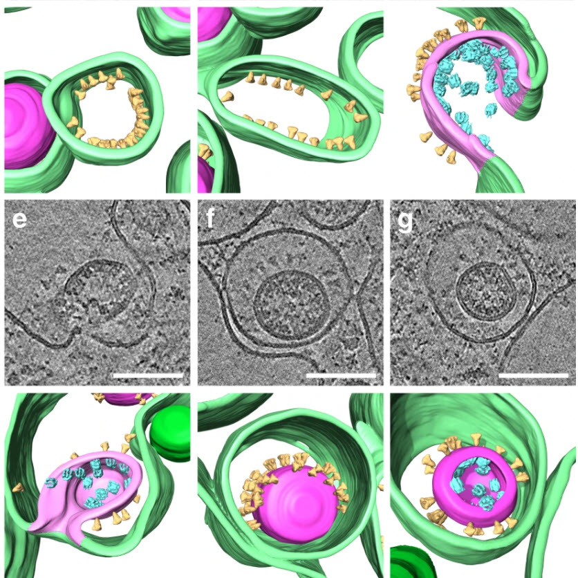
SARS-CoV-2 structure and replication characterized by in situ cryo-electron tomography
Severe acute respiratory syndrome coronavirus 2 (SARS-CoV-2), the causative agent of the COVID19 pandemic, is a highly pathogenic β-coronavirus. As other coronaviruses, SARS-CoV-2 is enveloped, replicates in the cytoplasm and assembles at intracellular membranes. Here, we structurally characterize the viral replication compartment and report critical insights into the budding mechanism of the virus, and the structure of extracellular virions close to their native state by in situ cryo-electr... Read more
Steffen Klein, Mirko Cortese, Sophie L. Winter, Moritz Wachsmuth-Melm, Christopher J. Neufeldt, Berati Cerikan, Megan L. Stanifer, Steeve Boulant, Ralf Bartenschlager, Petr Chlanda
STIM1 promotes migration, phagosomal maturation and antigen cross-presentation in dendritic cells
Antigen cross-presentation by dendritic cells (DC) stimulates cytotoxic T cell activation to promote immunity to intracellular pathogens, viruses and cancer. Phagocytosed antigens generate potent T cell responses, but the signalling and trafficking pathways regulating their cross-presentation are unclear. Here, we show that ablation of the store-operated-Ca2+-entry regulator STIM1 in mouse myeloid cells impairs cross-presentation and DC migration in vivo and in vitro. Stim1... Read more
Paula Nunes-Hasler, Sophia Maschalidi, Carla Lippens, Cyril Castelbou, Samuel Bouvet, Daniele Guido, Flavien Bermont, Esen Y. Bassoy, Nicolas Page, Doron Merkler, Stéphanie Hugues, Denis Martinvalet, Bénédicte Manoury & Nicolas Demaurex
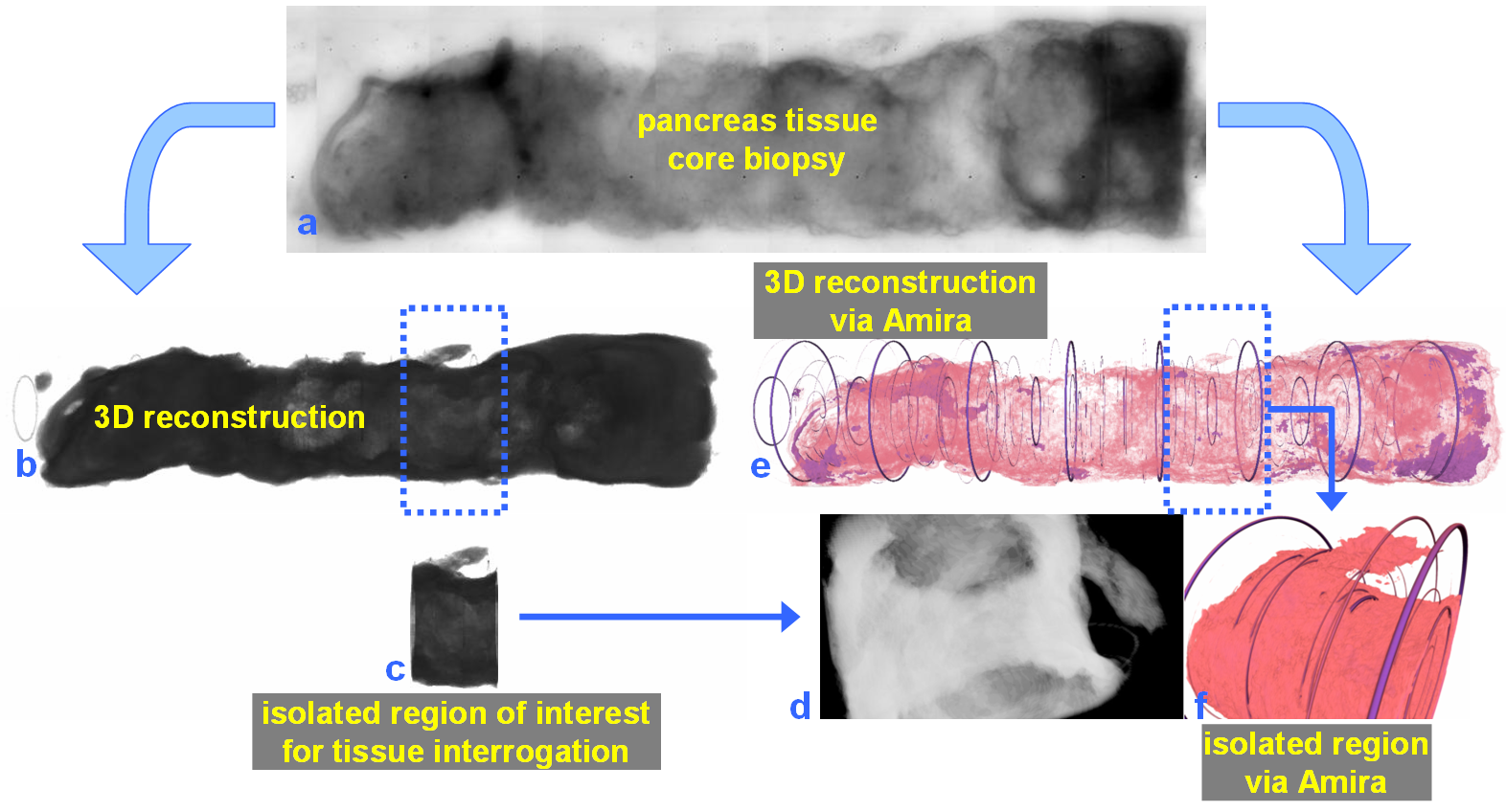
For nearly 100 years, pathology for cancer diagnosis has involved a standard, but complex series of steps to process tissue biopsies procured from a patient in the clinic. Many procedures are a direct result of the fact that observation and evaluation of specimens by pathologists occur using a standard microscope (in 2D).
In 2014, the Human Photonics Laboratory at the University of Washington demonstrated that the rudimentary operations of a pathology laboratory may be replicated on wh... Read more
Ronnie Das, PhD and Eric J. Seibel, PhD